Introducing Etch a Cell – Demolition Squad
Earlier this year, the team behind the Etch A Cell series of citizen science projects launched their latest project; ‘Etch A Cell – Demolition Squad’. Through this project, researchers based at the Francis Crick Institute (London, UK) have teamed up with Zooniverse volunteers to study ‘lysosomes’.
What are lysosomes?
Lysosomes are balloon-shaped organelles contain a range of enzymes that can break down biological substances including proteins, sugars, and fats. These enzymes allow lysosomes to function as the cell’s digestive system; lysosomes digest and degrade substances from both inside and outside of cells. This versatile functionality allows lysosomes to contribute to a range of biological tasks, including the breakdown of aged cellular components, the destruction of viruses, and the support of digestion during times of hunger. Lysosomes even play a role in the life cycle of cells by facilitating the removal of cells that have reached their expiry date. Etch A Cell – Demolition Squad has the aim of improving our ability to study lysosomes, to enable us to further understand how they contribute to the world of cellular dynamics.
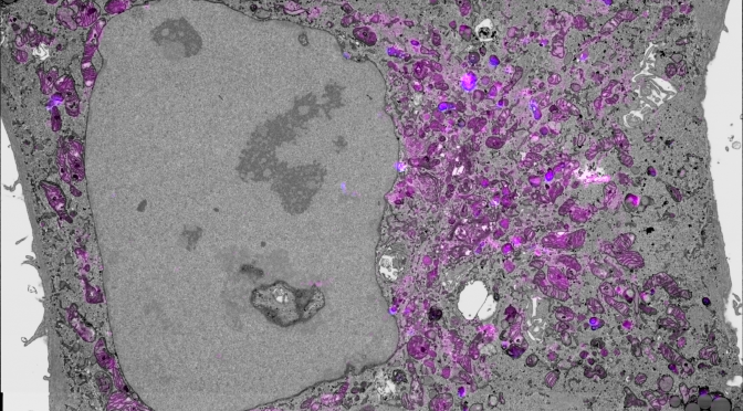
An image from the first data set analysed in Etch A Cell – Demolition Squad.
What’s the aim of the project?
The team behind Etch A Cell work with a variety of research teams to study different aspects of biology using cutting-edge Electron Microscopes. With their remarkable magnification and resolution, these microscopes allow researchers to capture intricate images of tissues, cells, and molecules. These images can be used to provide us with a richer understanding of biology, which can help us understand the biological changes associated with health and disease. Recent developments in electron microscope technology have enabled automatic image collection, leading to a deluge of data. This influx of information is helping propel research forward, however, it has caused a bottleneck in data analysis pipelines – which is why Etch A Cell needs the help of Zooniverse volunteers!
How are Zooniverse volunteers helping?
To study the images generated by our microscopes we typically analyse them by ‘segmenting’ the features we’re interested in, which means to draw around the bits of the cell that we want to examine. In Etch A Cell projects, Zooniverse volunteers help with this segmentation task. In Etch A Cell – Demolition Squad volunteers were asked to help with segmenting lysosomes. These structures can be very difficult to spot, so an imaging technique was used that enables the selective labelling of the lysosomes with a marker that allows their easier identification. You can see an example image below – the marker is shown in the images as pink, so volunteers were asked to draw around the grey blobs labelled with pink. In the image below you can also see some green lines which show how one expert segmenter drew around the lysosomes in this image.
In Etch A Cell – Demolition Squad, volunteers were asked to draw around lysosomes. To make the lysosomes easier to spot they were labelled with a marker, that is shown in the image above as pink. This image also shows lysosome segmentations done by an expert in green.
What are the results so far?
Since Etch A Cell – Demolition Squad was launched in May 2023, this project has received more than 12,000 classifications from hundreds of Zooniverse volunteers! This has allowed all 792 images in the first project data set to be retired. We’ve now started taking a look at the data, and we’re really impressed with the segmentations submitted! Well done to everyone who has contributed!
Here’s a sneak peek at some of the data you’ve produced through your collective efforts:
Briefly, you can see from these images show that Zooniverse volunteers have done fantastic job at this challenging task: the collective volunteer segmentations (shown in green in panel F) correspond really well to the location of lysosomes in the image (shown in pink in image B). With thanks to Francis Crick Institute internship student Fatihat Ajayi for generating these images.
The images above show the raw data (A) and this data overlaid with the pink marker that highlights the lysosomes (B). Image B is the one Zooniverse volunteers would have seen in the project interface. All the volunteer segmentations submitted for this image are shown overlaid in image C. Image D also shows the volunteer segmentations together, but in this image the line drawings have been ‘filled in’ and stacked on top of each other to make it easier to see where the volunteers agree there is a lysosome (the more volunteers who annotated a region, the brighter white it shows). Image E shows where most of the volunteers agreed there were lysosomes in this image, and image F shows how this corresponds to the raw data image (A). From these images you can see how collectively, Zooniverse volunteers did a great job at drawing around the lysosomes.
What’s next for Etch A Cell – Demolition Squad?
In the near future, we will be uploading new batches of data that we will need your help to analyse. These new data sets may look slightly different, but the task off segmenting the lysosomes will remain the same.
This project is part of the Etch A Cell organisation
‘Etch A Cell – Demolition Squad’ is one of multiple projects produced by the Etch A Cell team and their collaborators to explore different aspects of cell biology. If you’d like to get involved in some of our other projects, you can find the other Etch A Cell projects on our organisation page here.
Thanks for contributing to Etch A Cell!


